The content of the article
A pregnant woman is responsible for two lives: her own and the developing embryo. Childbearing is a natural process, but complex and responsible, so the expectant mother is constantly worried and worried. How many babies grow inside, are there any pathologies and how is the fetus located? The doctor doing the ultrasound will soothe and answer all these questions. But how many procedures should there be? How harmful are they? Maybe it's better to do without ultrasound?

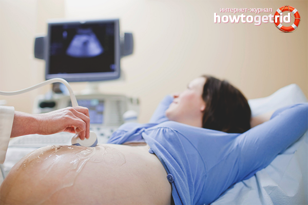
First trip to the diagnostician
The girl, who noticed symptoms of pregnancy in her or decided to confirm the test results, the gynecologist gives directions to a free ultrasound examination. You can also go through the procedure in a private clinic, if the queue is large, and you need to urgently find out whether the patient will become a mother.
A woman who came to the diagnostician at an early period is examined with a transvaginal method. The specialist uses a long hose equipped with a wave emitter. He inserts the device into the vagina, and thanks to this method, it accurately determines whether the fertilization of the egg has taken place, how large the embryo has grown, and how many weeks the fetus has grown. The first ultrasound is considered to be verifiable and unscheduled. The woman is informed of the news and given time to decide how much the child is desired, and how to proceed.
Trimester One: Embryo Formation
The patient discussed her condition with her husband or relatives, consulted with her friends, thought carefully about the pros and cons and realized that she had long wanted to try herself as a mother. It remains to be registered with the gynecologist and follow all instructions of the female doctor. The first is to undergo another ultrasound at 10–14 weeks.
Patients whose pregnancy was diagnosed at 2-3 months, you can refuse the procedure. Provided that the fetus did not find any abnormalities, the uterus is completely healthy, and the gynecologist did not suspect placental abruption.If the girl came to the reception by an ultrasound specialist at 4–8 weeks, you will have to go to the examination again.
Why go through the procedure in the first trimester? To become registered with a gynecologist, collect all information and get a pregnant card. During the first ultrasound, the diagnostician will tell you how many embryos develop in the womb, and whether there is a threat of miscarriage. At weeks 10–14, specialists find genetic abnormalities such as hydrocephalus and Down syndrome.
In the early period it is impossible to abandon the ultrasound, because thanks to this procedure, an ectopic pregnancy is determined and the patient's life is saved. The specialist will assess the condition of the growing fetus and tell you what date to give birth.
For a trimester, a healthy woman should undergo only one ultrasound. A pregnant woman can send a second consultation to a diagnostician if:
- uterine bleeding has opened;
- the gynecologist suspected that the fetus froze;
- the embryo grows slowly and does not meet the standards;
- a woman complains of lower abdominal pain;
- at the first examination, the diagnostician suspected placental abruption;
- In the early stages of the mother had to take illegal drugs or work with toxic substances.
If the gynecologist suggested to repeat the screening in a week or a few days and pass additional tests, you should not worry. Experts want to collect a detailed history of the mother and fetus to make a definitive diagnosis and refute any suspicions that have arisen.
Trimester Two: the baby continues to grow
A pregnant woman should undergo a second ultrasound scan at 20–24 weeks. The diagnostician will estimate how much the belly of the mother and the child developing inside have grown. At this time, screening is done to determine the pathology:

- hearts;
- nervous system;
- brain;
- back bifida;
- cleft lip;
- lack of brain;
- wolf's mouth.
The second wave study determines the syndromes of Patau and Edwards, as well as the gender of the future baby. You can invite the father of the child with you to find out who will be born: daughter or son. Doctors at the 24th week conduct an ultrasound examination of the vessels of the placenta to prevent preeclampsia. Pregnant women should visit the diagnostician in time, follow the recommendations of the gynecologist and not be afraid to undergo additional ultrasound.
Third Term: Final Conclusions
In the period from 32 to 34 weeks, the woman will have the last scheduled screening. Diagnosis allows you to identify the child malformations that can be surgically removed. The fetus will be operated on in the womb, and the baby will be born completely healthy.
Thanks to ultrasound, doctors will determine the weight and size of the child, and then decide which method of birth to choose: natural or cesarean section. The diagnostician evaluates:
- fetal position: head to the left or right side, up or down;
- condition of the uterus and birth canal;
- whether the umbilical cord wraps around the neck or body of a child.
Additionally compare the size of the birth canal and the volume of the head of the fetus. If the baby seems too large, a woman will be offered a caesarean section. Routine surgery will help to avoid serious injuries and heavy blood loss.
Ultrasound in the third trimester is necessary for doctors to work out the tactics of childbirth. In addition to screening in a child, the pulse is measured and a cardiogram is made, and the circulatory system is examined. An additional ultrasound can be prescribed to a pregnant woman immediately before delivery, if a previous examination revealed cord entanglement, improper positioning of the baby or problems with the mother.
Reasons for additional research
A healthy mother for all nine months should visit the diagnostic room from 3 to 5 times. It is safe and does not harm the baby or the woman herself. Ultrasonic waves are emitted at a frequency of 2 to 10 MHz, fighting off the soft tissue of the fetus. The device does not irradiate the child and does not cause the development of pathologies.
You can undergo an ultrasound at least every 2–3 weeks, if the gynecologist thinks there is a need. Pregnant women can become regular visitors to the diagnostic room:
- there was a rubella during gestation;
- previous pregnancy ended in miscarriage or the birth of a dead baby;
- the baby's father is a close relative;
- family history has hereditary diseases;
- rhesus conflict with the baby;
- chronic diseases can threaten the life of a woman or fetus.
Additional studies are conducted when a pregnant woman expects double or triplet, or all of the symptoms indicate a placental abruption. The patient may also be recommended to undergo an unplanned dopplerography if she has changed blood clotting or has started preeclampsia.
3D study
In conventional clinics used two-dimensional scanning of the fetus, which allows us to consider the anomalies and monitor the development of the child. In some modern centers, future mothers offer a new service - three-dimensional ultrasound.
This procedure should be performed only from 20–21 weeks. The number of scans may be unlimited, but it is desirable to resort to this type of research only in cases where there are serious reasons for it:
- suspicion of an abnormality or disease that cannot detect a two-dimensional scan;
- fertilization occurred artificially with the help of IVF;
- the woman decided to become a surrogate mother;
- gestation is accompanied by serious complications.
Numerous studies have shown that ultrasound does not lead to fetal abnormalities. But the procedure, like taking pills or a vaccine, is not completely harmless. There is no need to refuse screening, but one should not make additional diagnostics without the recommendation of a gynecologist. You can not abuse the three-dimensional and four-dimensional scans, because they use more powerful radiation.
Ultrasound is an important and necessary procedure that should not be neglected by a pregnant woman. It is painless and safe, helps to control the development of the fetus. Over the entire period of gestation, you can make from 3-5 scans to 10-30, if there is a threat of miscarriage or complications. But do not be afraid that it will harm the baby. No, ultrasound will help make and give birth to a healthy baby, who will please his mother every day.
Video: how often during pregnancy you can do an ultrasound


To send